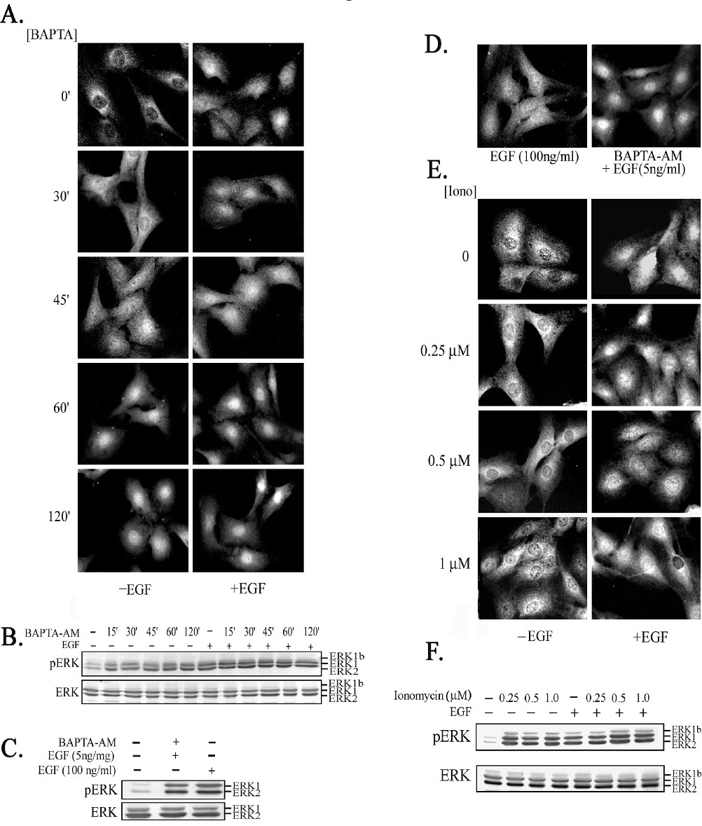
Fig. 4. Subcellular localization of ERKs in response to variable calcium concentrations. A. Following serum-starvation (0.1% FBS, 16 h), Rat1 cells were treated with BAPTA-AM (15 µM) for varying times, with EGF (50 ng/ml for 15 min) or pretreated with BAPTA-AM prior to EGF stimulation for the final 15 min of the treatment. The cells were stained with anti-ERK Ab and its localization was assessed by fluorescence microscopy. B. Cell lysates were subjected to SDS-PAGE and Western blot analysis using Abs to pERK (upper panel) or gERK (lower panel). C. Rat1 cells were treated with 15 µM BAPTA-AM for 1 h prior to stimulation with 5 ng/ml EGF or with 100 ng/ml EGF alone. Lysates were subjected to Western blotting with anti-pERK Ab. D. Rat1 cells were treated with 100 ng/ml of EGF for 15 min, BAPTA-AM for 60 min with 5 ng/ml EGF 15 min and the localization of ERK was assessed with anti-gERK Ab. E. Rat1 cells were treated for 30 min with varying concentrations of ionomycin alone, or prior to EGF stimulation (50 ng/ml, 15 min). The localization of ERK was assessed with anti-gERK Ab F. Cell lysates were subjected to Western blot analysis using anti-pERK (upper panel) or gERK (lower panel) Abs.